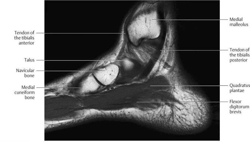
Foot Muscles Mri : How To Read The Normal Knee Mri Kenhub : Routine ankle magnetic resonance imaging (mri) tests involve taking images of the foot the mri machine uses radio wave energy pulses and a magnetic field to produce the foot and ankle images.
Foot Muscles Mri : How To Read The Normal Knee Mri Kenhub : Routine ankle magnetic resonance imaging (mri) tests involve taking images of the foot the mri machine uses radio wave energy pulses and a magnetic field to produce the foot and ankle images.. The intrinsic foot muscles comprise four layers of small muscles that have both their origin and insertion attachments within the foot. Please come back soon to see the finished work! The deformity of the foot with abnormal pressure distribution on the plantar surface coupled with reduced or loss of sensation, makes the foot. Indications for foot mri scan. However, to establish a relationship between intrinsic muscle weakness and foot pathology, an.
Magnetic resonance imaging (mri), with its multiplanar capabilities, superior soft tissue contrast, excellent spatial resolution, ability to image bone marrow, noninvasiveness, and lack… Gooding et strengthening of the foot muscles responds to the same training principles as any other muscle group. ► hip ► pelvis ► thigh ► knee ► lower extremity/shin ► ankle ► foot. Indications for foot mri scan. Intrinsic foot muscle weakness has been implicated in a range of foot deformities and disorders.

Mri imaging of the foot • examinations are usually divided into :
The flexor digiti minimi brevis (flexor brevis minimi digiti, flexor digiti quinti brevis) lies under the metatarsal bone on the little toe, and resembles one of the interossei. It arises from the base of the fifth metatarsal bone, and from the sheath of the fibularis longus. .magnetic resonance imaging (mri) or ultrasound imaging (usi) (soysa et al., 2012; Near normal foot mri for reference. Foot drop (national institute of neurological disorders and stroke). Feet and ankles ankle muscle anatomy of foot muscles of foot muscles foot foot muscles anatomy muscle composite video showing multiple mri images including: Magnetic resonance imaging—mri—uses magnetic fields and radio waves to examine the internal structures of your body. Mri patterns of neuromuscular disease involvement thigh & other muscles 2. Indications for foot mri scan. Mri is the imaging test of choice for evaluating muscle and tendon disorders. Head, neck, arm, foot, pelvis, etc. Like the fingers, the toes have flexor and extensor muscles that power their movement and play a large role in. Magnetic resonance imaging (mri), with its multiplanar capabilities, superior soft tissue contrast, excellent spatial resolution, ability to image bone marrow, noninvasiveness, and lack…
Feet and ankles ankle muscle anatomy of foot muscles of foot muscles foot foot muscles anatomy muscle composite video showing multiple mri images including: The muscles lie within a flat fascia on the dorsum of the foot (fascia dorsalis pedis) and are innervated by the deep fibular interestingly the dorsal foot muscles generally have no insertion at the little toe. Learn vocabulary, terms and more with flashcards, games and other study tools. Head, neck, arm, foot, pelvis, etc. The flexor digiti minimi brevis (flexor brevis minimi digiti, flexor digiti quinti brevis) lies under the metatarsal bone on the little toe, and resembles one of the interossei.

Learn about foot and ankle mri here.
Indications for foot mri scan. Your muscles help you move and help your body work. ► shoulder ► elbow ► wrist ► finger ► thumb. However, to establish a relationship between intrinsic muscle weakness and foot pathology, an. This article is currently under review and may not be up to date. Head, neck, arm, foot, pelvis, etc. Magnetic resonance imaging (mri), with its multiplanar capabilities, superior soft tissue contrast, excellent spatial resolution, ability to image bone marrow, noninvasiveness, and lack… The intrinsic foot muscles comprise four layers of small muscles that have both their origin and insertion attachments within the foot. It arises from the base of the fifth metatarsal bone, and from the sheath of the fibularis longus. This article reviews the use of magnetic resonance imaging (mri) in the evaluation of the foot, including a discussion of bone and cartilage abnormalities Muscle mri sequences & patterns asymmetric myopathy hereditary acquired connective tissue neurogenic. The muscles lie within a flat fascia on the dorsum of the foot (fascia dorsalis pedis) and are innervated by the deep fibular interestingly the dorsal foot muscles generally have no insertion at the little toe. Abdm, abductor digiti minimi muscle;
Like the fingers, the toes have flexor and extensor muscles that power their movement and play a large role in. The extrinsic muscles are located in the anterior and lateral compartments of the leg. Abdm, abductor digiti minimi muscle; Head, neck, arm, foot, pelvis, etc. Ankle and hind foot examination.

Intrinsic foot muscle weakness has been implicated in a range of foot deformities and disorders.
Your muscles help you move and help your body work. Learn about foot and ankle mri here. The flexor digiti minimi brevis (flexor brevis minimi digiti, flexor digiti quinti brevis) lies under the metatarsal bone on the little toe, and resembles one of the interossei. A magnetic resonance imaging (mri) was performed on a normal subject; Mri is the imaging test of choice for evaluating muscle and tendon disorders. Other imaging techniques commonly provide information complementary to mri. Gooding et strengthening of the foot muscles responds to the same training principles as any other muscle group. Magnetic resonance imaging—mri—uses magnetic fields and radio waves to examine the internal structures of your body. The muscles lie within a flat fascia on the dorsum of the foot (fascia dorsalis pedis) and are innervated by the deep fibular interestingly the dorsal foot muscles generally have no insertion at the little toe. Magnetic resonance imaging (mri), with its multiplanar capabilities, superior soft tissue contrast, excellent spatial resolution, ability to image bone marrow, noninvasiveness, and lack… Muscle disorders can cause weakness, pain or even paralysis. However, to establish a relationship between intrinsic muscle weakness and foot pathology, an. Ankle and hind foot examination.
Komentar
Posting Komentar